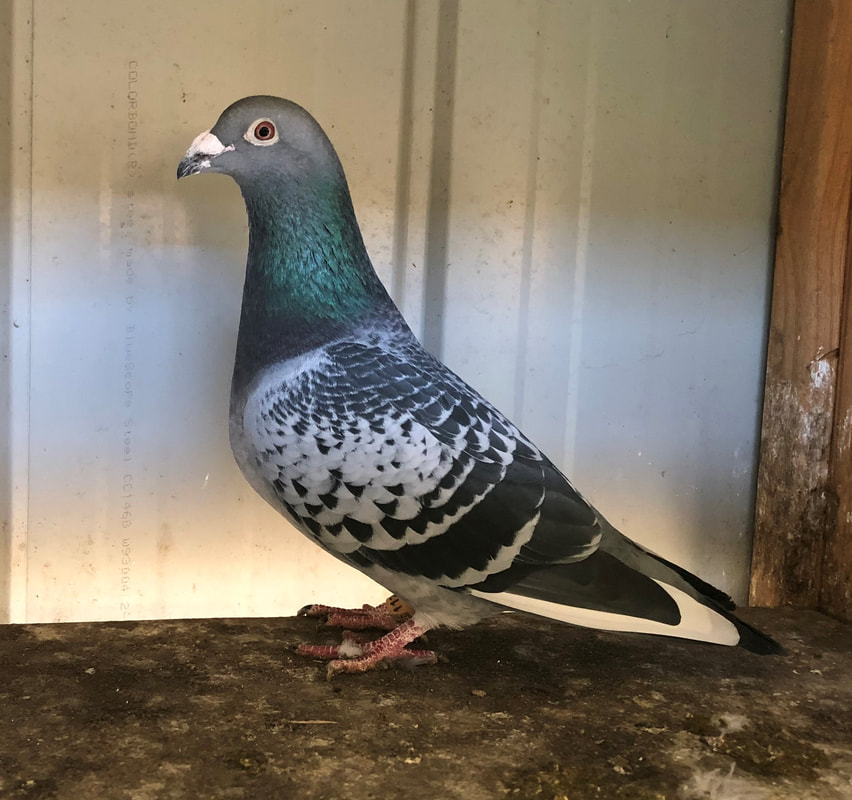
VHA 17 11220, 2nd VHA Federation for Colin Walker. Rankin Springs, 490 km, 895mpm, 1/9/18
|
The Flying Vet - Veterinary Updates |
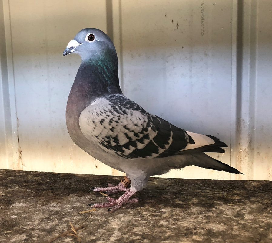
VHA 17 11348 BCPH , 1st VHA Federation for Colin Walker, Griffith, 418km, 1132mpm, 21/10/18
|
|
Looking back, it is interesting to note how veterinary pigeon matters have changed over the last 40 years but particularly during the last 10 years. The level of diagnostic ability and veterinary knowledge has dramatically increased and along with this fanciers’ expectations from avian veterinarians have also increased. There is, however, still a surprising amount of misinformation about. Some fanciers are real “gossips”, others are “wannabe” vets and sometimes it seems as if everyone is a veterinary expert. Unfortunately, not all misinformation originates from fanciers, sometimes it is from a vet. Like all professions, the veterinary profession has one or two rogues. Most vets are informed and dedicated to their profession but sadly some are less dedicated and some could even be described as crooks or charlatans. In the past, there were only a small number of qualified avian vets and some fanciers, when they think of avian vets, still think that there are only one or two who can help them with a health problem with their pigeons. There are now, however, about 50 qualified avian vets in Australia. Not all are familiar with pigeon racing but all are familiar with pigeon health problems, the appropriate diagnostic process to investigate these and the correct use of medication. They are able to diagnose medical problems ranging from the minor ones that make race birds a bit less competitive through to the more serious ones where birds are dying. Once a diagnosis has been made, they can then advise a fancier on how the problem developed, how to control it and how to stop it happening again.
At the Melbourne Bird Veterinary Clinic (MBVC), there are four veterinarians and three of these are qualified avian vets. When dealing with a health issue, it is always a matter of correlating the degree of diagnostic effort and expense with the severity of the problem. If the birds are racing well and we are simply monitoring health, then just a general examination and a microscopic examination of a crop flush and faecal smear may be all that is required. Other tests can be done if indicated after examination. With more serious problems, the following is the diagnostic protocol that is followed at the MBVC. This is typical of a thorough diagnostic investigation in most avian practices and can be used as a guide for your veterinarian. The 12 step process to investigate a serious pigeon health problem 1/ Take a history and develop an understanding of the nature of the problem. 2/Conduct a thorough clinical exam, make assessment of general clinical condition, undertake a microscopic exam of fresh faecal smear, conduct a faecal flotation, undertake a microscopic exam of fresh crop aspirate, check pharyngeal tonsils and laryngeal mound (redness, swelling, abscessation, sialoliths, etc.) in particular. 3/Collect throat, conjunctival (eye) and choanal (‘slot’) swabs for Chlamydia and Mycoplasmal PCRs. 4/Collect blood for full haematology and biochemistry – this gives valuable information about nutrition, the level of training and overall fitness, hydration, organ function, the immune system and infection. This information cannot be gathered from autopsy. 5/ Euthanise bird by giving lethal injection; usually pigeons just “fall asleep”. 6/ Wait 2 hours to do the autopsy; earlier leads to passive bleeding, identified as “congestion” by pathologists, which can make interpretation of tissues microscopically difficult or impossible. 7/ Autopsy within 4 hours of death, later can lead to early autolytic (decomposition) changes and altered bacterial populations, decreasing diagnostic value. 8/ Collect full set of tissues including -- whole head (after removal make a longitudinal incision along the top and back of the skull and crack the skull open to allow formalin to run into the brain case), the anterior oesophagus (sometimes this is the only place you will find Herpes), sections of crop wall, proventriculus, gizzard, several sections of gut, caeca (vestigial) and bursa of Fabricius if visible (i.e. bird <5 months old), trachea, syrinx, lungs, air sac, spleen (often best to collect on first opening the abdomen, if there is bleeding spleen can later be hard to find), liver (left and right lobes), kidney, middle-third of femur (for bone marrow), sciatic nerve, section of spine, skin, heart and pectoral muscle. 9/ Collect PCR for Rota in case you get hepatocellular necrosis of unknown cause; does not matter if the bird is vaccinated against Rota as the PCR does not check for the tiny part of the genome that codes for the capsid VP8 protein that forms the basis of the subunit vaccine. Birds can be negative for Rota on faecal PCR and positive on hepatic PCR. Collect kidney and bowel PCRs for PMV PCR testing. 10/ Collect a second set of the main tissues and freeze these. These are used if further viral identification and testing are required 11/ If anything looks abnormal, target that and make sure a section/sample is collected. 12/ If anything looks infected, collect a swab for MC and S (microscopic examination, culture and antibiotic sensitivity) testing. Collect a similar swab of the gut content. Everything costs money. It is important that the client’s money is spent wisely and so the samples collected in points 3, 9, 10 and 12 above are held and available if the other tests, in particular the blood profiling and histopathology, are not diagnostic. These samples cost nothing to collect, store well and some can only be collected from a dead pigeon. Overlooking the collection of these may not only compromise the diagnostic process but may necessitate killing another bird unnecessarily to subsequently collect these samples. Sometimes fanciers have an unrealistic expectation as to what can be diagnosed by fairly basic tests. Sometimes vets are overly keen to give fanciers a diagnosis and make impossible diagnostic claims after inadequate sampling and testing. From a mailed in dropping sample it is only possible to definitively diagnose (and even then, not always) some worms and coccidia. It is not possible to diagnose (or rule out) bacterial or fungal infections. In particular, Streptococcus and Salmonella infections ( just because the organism may be present in the droppings does not mean it is causing disease ) cannot be diagnosed this way. To consider diagnosing any form of canker or respiratory infection in this way is just ridiculous. Any vet who claims to make this diagnosis in this way is deluding not only the fancier but also himself. Similarly it is not possible to diagnose bacterial or fungal infections from a mailed-in throat swab. I am often asked to review cases and offer a second opinion. Most vets do really great work but the standard of work of some is well below the average of what the profession would expect. A recent case illustrates the point. A fancier was experiencing both poor results and poor returns in his race team. He went to the trouble and expense of sending a live pigeon to an avian vet interstate for diagnostic investigation. On arrival, the bird was handled and droppings were looked at under the microscope. The bird was then euthanized, autopsied and some tissue samples collected for histology (microscopic examination). The pathologist reported that many of the samples were “congested”. From this diagnostic work, the vet was happy to make a diagnosis! He diagnosed that the bird had come in contact with a toxin and asked the fancier to send some grain for testing. The grain was placed onto a medium that stimulates bacteria and fungus on the surface of the grain to germinate. This test is not regarded as an accurate way of assessing grain quality or an accurate measure of fungal or bacterial contamination. But from this test, the vet advised the fancier that the corn was a problem. The fancier stopped using the corn. Nothing changed and the fancier then re-contacted the vet who advised that it was a “very complex” problem and another bird would need to be sent for testing. The fancier then contacted the MBVC. The vet had not necessarily done anything wrong but most vets would regard his diagnostic investigation as pretty poor. Comparing his work with the above recommended 12-point protocol, many problems are apparent. The main ones are listed below. Problems with this investigation a/ Inadequate collection of samples required for an accurate diagnosis. When asked why various samples were not collected, his reply was that “he would rely on the histopathology” for a diagnosis. A seriously flawed approach with many potentially relevant and important things being overlooked. b/ Poor autopsy technique – when asked if the “congestion” noted by the pathologist was apparent at autopsy or if he had caused it, his simple reply was “I don’t know”. Many important tissue samples were not collected during the autopsy. Yet, despite inadequate tissue collection and poor tissue handling, this was his main diagnostic test. c/ His preparedness to make a diagnosis that was impossible based on the test results. What concerned me is that when I contacted the vet in question, he thought he had done a good job. Unfortunately, he had become professionally isolated and his knowledge and diagnostic approach were no longer in step with the profession’s expectations. The frustrating thing for me was that he did not seem to realise this. He seemed simply out of touch with the diagnostic process and also with the standard way that most avian vets would approach this problem. On top of this, some of his testing and advice was flawed and often just wrong and certainly not consistent with what would be regarded as acceptable practice in the profession generally. Veterinarians as professionals may differ in the exact way that they diagnose and manage cases but in this instance the tests being done and treatment recommended did not have a scientific basis. He was substituting his opinion for scientific fact. I found it particularly interesting that he was quick to label this a “complex case”. These days, nothing should really be complex given the correct testing. These days, diagnostic testing is so sophisticated that it is simply a matter of following the usual diagnostic pathway and getting the results. And so what was wrong with this fancier’s birds? The haematology component of the blood profiling revealed a very high PCV. This stands for Packed Cell Volume and is an indicator of the concentration of red blood cells in the circulation. When PCVs get high, the blood becomes more viscous and the heart has to work harder to pump it around the body. This puts a dramatic increase on the work load of the heart and leads to cardiac insufficiency during exertion and an obvious compromise of race performance. This condition is seen quite commonly. The exact cause of these high PCVs is not known but one suggestion is that it is the response of a pigeon being forced to exercise beyond its fitness or health capability. The condition usually responds to treating any identified health problems ( that are blocking the development of fitness) and simply to just open lofting. Affected birds are kept in their loft routine, i.e. let out and called in at their normal times but are not forced to fly. Leaving birds at liberty for several hours a day I find is a good stress reliever and helps the birds to gradually fly themselves to fitness – those that want to fly do and those that want to rest can, as they start to feel well they fly more and develop their own fitness. During this time, there is no rationing of food. Birds are fed by hand as much as they want to eat twice daily with a moderate protein ration of about 16%, e.g. 40% dun peas, equal parts (about 15% of each) safflower, sorghum, wheat and corn. Usually the condition corrects itself over 2 weeks and the team can gradually be brought back into work. The biochemistry component of the blood profile showed minor damage to the liver and reduced kidney function but histopathology showed no significant damage at a cellular level and subsequent Rota and PMV PCRs (viral DNA checks) ruled out these viruses as being involved. All other tests did not reveal any other complicating factors. There was no evidence of a “toxic” exposure. Moving on from this I have even reviewed cases where vets have invented conditions and then advertised that they are the only ones to be able to diagnose that condition and in turn, market elaborate treatment programs to manage the condition. A list of qualified avian vets in Australia and New Zealand is listed on the Australian Pigeon Company website. I would encourage all fanciers to use their services but at the same time, if the advice you are getting just does not sound right, get a second opinion.
1 Comment
2/28/2020 03:22:05 am
Very interesting update on your blog thank you so much for sharing. The information here about the pigeon here would be effective and the people who are having them they able to explore as well. Also we able to diagnose the problem with our pets as well through the information. Leave a Reply. |
AuthorDr Colin Walker Archives
October 2020
Categories
All
|
 RSS Feed